
Mammogram showing the presence of a breast cancer (red arrow)
What are the treatments for breast cancer?
Breast cancer management consists of a multimodality treatment, which means that different specialists use a variety of treatments to treat the same cancer. The broad categories are:
Breast cancer surgery, which Dr Bell will perform
Radiation treatment (Radiotherapy), under the care of a Radiation Oncologist
Systemic treatment (including hormonal therapy, chemotherapy and Herceptin administration), under the care of a Medical Oncologist
All these specialists are part of a multidisciplinary team that comes together to optimise your individual cancer treatment pathway.
There is a lot of research happening in the field of breast cancer, which means that we can continue to offer better treatments and improve the survival rates amongst breast cancer patients.
In what order are the breast cancer treatments performed?
Breast cancer surgery is usually the first treatment, whilst the other treatments are known as adjuvant treatments (additional treatments to surgery). However, sometimes we recommend starting breast cancer treatment with chemotherapy, followed by surgery. This is referred to as neoadjuvant chemotherapy.
What are the surgical aims of breast cancer treatment?
There are 2 surgical aims for breast cancer surgery:
To remove the breast cancer with a rim of normal breast tissue (clear margins). This can either be performed with breast conserving surgery or a mastectomy.
To find out if the cancer has spread to the lymph nodes (stage the axilla). This will either be apparent on pre-operative imaging and confirmed with a biopsy or assessed at the time of surgery with a sentinel lymph node biopsy.
What are the types of breast surgery for breast cancer?
Breast Conserving Surgery (including Oncoplastic Breast Surgery) +
Breast Conserving Surgery
This is also known as a “lumpectomy” or “wide local excision” (WLE). This procedure involves removing the breast cancer with a rim of normal breast tissue (clear margins). It always requires adjuvant radiotherapy to the rest of the breast, to reduce the chances of tumour recurrence.
If the tumour is not able to be felt, it will require localisation on the day of surgery with a hookwire. This is a thin wire that gets inserted into the breast (under guidance of breast imaging) by a radiologist in the radiology department. Dr Bell will use this wire during the operation to guide the excision of the abnormal breast tissue.
The removed breast tissue will be sent for imaging (specimen Xray) to ensure the abnormality is contained within it and subsequently to the pathology lab for a final result.
Occasionally, despite the imaging showing an apparent margin of normal tissue around the tumour, the pathology report demonstrates involved margins with the tumour cells. In this instance a further procedure is required (re-excision of the margins) to obtain clear margins.
The aim of the procedure is to remove all abnormal tissue, but to maintain the shape and appearance of the breast as much as possible. The larger the tumour, the more difficult this becomes. For patients which larger tumours, sometimes breast conserving surgery is still possible with the help of oncoplastic techniques.

Image 1. Mammogram with 2 hookwires localising an area of abnormal calcification (green circle). Image 2. Specimen X-ray showing the presence of the calcification in the excised breast tissue.
Oncoplastic Breast Surgery
Oncoplastic refers to using plastic surgery techniques to close the cavities (defects) left in the breast after removal of the cancer. These techniques are useful after removal of larger cancers and consist of remodelling the breast tissue by shifting the breast tissue around and creating a smaller overall breast size, whilst maintaining the breast contour (similar to techniques used in breast reduction surgery). Other techniques include replacing the removed breast tissue with tissue from outside the breast itself (flaps).
Oncoplastic breast surgery is more technically challenging and takes longer than traditional breast surgery. In addition, it often requires symmetrising surgery to the opposite breast. Not every patient needs or is suitable for oncoplastic breast surgery. Dr Bell has undergone additional training in oncoplastic breast surgery during her Post Fellowship Training (PFT) via BreastSurgANZ.
Mastectomy +
A mastectomy involves removing all breast tissue including the nipple and surrounding skin and leaving a single scar across the chest. Ideally, a flat chest wall is achieved, that will support a breast prosthesis in a bra or will be suitable for breast reconstruction in the future.
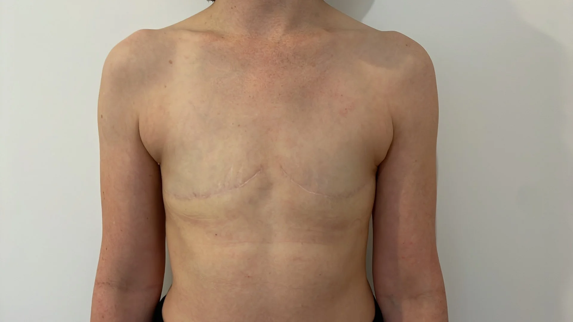
Photo of a patient post bilateral mastectomy by Dr Bell
(posted with permission)

What are the risks of breast cancer surgery?
Breast cancer surgery is usually well tolerated, and most patients can go home the next day. Interestingly, the majority of patients report these procedures to be less painful than might be expected. The general risks include infection, wound problems, bleeding (including a blood collection/haematoma that might require further treatment in theatre) and fluid collections (seromas). In breast conserving surgery, occasionally further surgery is required to obtain clear margins. In a mastectomy, a drain will be left during the operation to drain excess fluid from underneath the wound. Once drain volumes decrease, the drain will be removed. However, fluid can continue to build up and may require subsequent needle drainage in the rooms. Breast cancer surgery can also influence your psychosexual wellbeing. Dr Bell will address this issue and discuss any concerns you may have.
How do I know if the breast cancer has spread to my lymph nodes?
If breast cancer spreads beyond the breast, it tends to go the lymph nodes first and can subsequently spread to the rest of the body. If this happens, most commonly, breast cancer will go the lymph nodes in the armpit (axillary lymph nodes) on the same side as the breast cancer. Less common, it can spread to lymph nodes deep to the ribs, located next to the breastbone (internal mammary lymph nodes). At the time of your breast imaging, your axillary lymph nodes will be checked with an ultrasound. If they look abnormal, a biopsy may be performed to check for the presence of cancer cells. Dr Bell will discuss the appropriate lymph node surgery with you. Generally, most patients with “normal” appearing lymph nodes will be suitable for a sentinel lymph nodes biopsy and patients with cancer in the lymph nodes will be offered an axillary lymph node dissection.